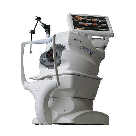
Optical Coherence Tomography 3D OCT-1 Maestro
Product Overview
Full-auto Capturing
3D OCT-1 Maestro requires nothing more than to touch the capture icon and [Start Capture] button. Alignment, focus, optimizing, and capturing are performed in automatic procedure. After the capturing, report can be immediately displayed by clicking on the icon.

Semi-auto Capturing
With semi-auto capturing, 3D OCT-1 Maestro completes alignment, focus and optimizing automatically, then allows for an operator to start capturing at any convenience. This enables to easily find the best timing to capture with communicating with patients even in difficult cases.

Ease of Use for Capturing Small Pupils
OCT-LFV image, which is a live projection image with reflection at retina, will show the live Fundus image clearly even in cases of smal pupilis. Disc, retinal vesseles and scanning position is very easy to see.
Complete OCT Functionalities
- For Macula
– 5 Line Cross Scan
– 3D Macula Analysis
– Line Scan
– Radial Scan
- For Glaucoma
– 3D Wide Scan (12mm × 9mm)
– 3D Macula(V) Glaucoma Analysis
– 3D Disc Analysis
 3D Wide Scan(12mm × 9mm) 3D Wide Scan(12mm × 9mm) |
 Trend Analysis(GCL) Trend Analysis(GCL) |
High Quality / High Resolution OCT and Color Fundus Photography
50,000 A-scans/sec. speed produces fine B scan and smooth 3D graphics, which facilitates the observation of pathology form and condition on each layers. High quality color fundus photography gives fundamental and additional information. The OCT and color fundus can be said to be the inseparable combination for daily diagnosis.


- Fully-automatic OCT with simple finger touch
- Rich analysis and report functions
- Reliable assistance for scanning
- High quality, high resolution OCT and color fundus image
- Seamless network solution
- Compact footprint and flexible layout

